
Types, Safety, Benefits, and When They're Needed
Author; S.Raj, M.D, Updated: 7/22/2026 Reading Time: 5 Minutes
Dental X-Rays and CBCT Scans Explained
Imagine visiting the dentist with what feels like a healthy smile—no pain, no visible cavities, and your teeth and gums seem perfectly fine. Yet beneath the surface, issues like hidden decay, jaw cysts, or impacted teeth could be quietly advancing, invisible during a regular dental exam. That's where dental x-rays and advanced 3D scans step in—not as routine checkboxes, but as vital, carefully chosen tools for accurate diagnosis and truly individualized care.
Accurate diagnosis matters. Dentists want to catch problems early and design a treatment plan just for you. Sometimes, a simple mirror check isn’t enough. Some dental conditions are hidden from view and may go unnoticed during a visual dental exam. Dental X-rays and CBCT scans enable dentists to detect cavities, bone loss, infections, and impacted teeth that can’t be seen otherwise. Imaging helps ensure that nothing critical is overlooked and that each recommendation fits your unique needs.

Dentists use dental X-rays only when the expected benefit to oral health is greater than any potential risk.
Dental X-rays and CBCT scans provide valuable diagnostic information that helps dentists plan safe, accurate, and personalized treatment while using imaging only when clinically justified.
What Are Dental X-Rays?
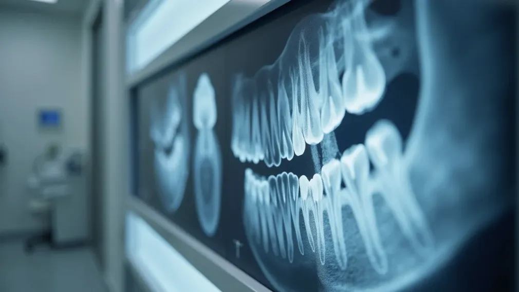
Dental X-rays, also known as dental radiographs, are special medical images that use low doses of ionizing radiation to help your dentist examine the teeth, jawbone, and oral structures from the inside, not just the outside. While your visual dental exam checks the surface, dental X-rays reveal what’s hiding between, under, and inside the teeth and jaws—where cavities, infections, cysts, and bone loss may develop.
Dental X-rays, also called dental radiographs, are medical images that use low levels of radiation to help dentists examine teeth, bone, and surrounding oral structures.
Dental X-rays show details like tooth decay, bone health, infections, and abnormalities that a dentist cannot see with the naked eye.
Most dental X-rays create 2-dimensional images—a "flat" view of the area of interest. This is often sufficient to diagnose and treat common dental problems, such as decay between your back teeth or bone loss from gum disease. However, some issues may be difficult to spot or fully evaluate with 2D images alone. This is when your dentist might recommend advanced 3D dental imaging, like a CBCT scan, to get a clearer, more complete picture before proposing a treatment plan. All dental imaging is performed when there's a clear benefit for you, and your dentist will always explain why a certain radiographic examination is needed.
For those interested in how dental imaging fits into the broader landscape of oral healthcare, exploring the range of dental services available can provide additional context on how local practices utilize advanced diagnostic tools to enhance patient outcomes.
Types of Dental Imaging: Bitewing, Periapical, Panoramic X-Rays and CBCT Scan
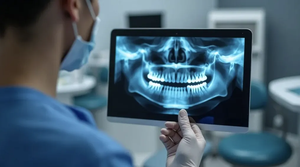
Not all dental X-rays are the same. Dentists select from several types of dental radiographs to target different parts of your mouth and answer specific diagnostic questions. Here are the most common types:
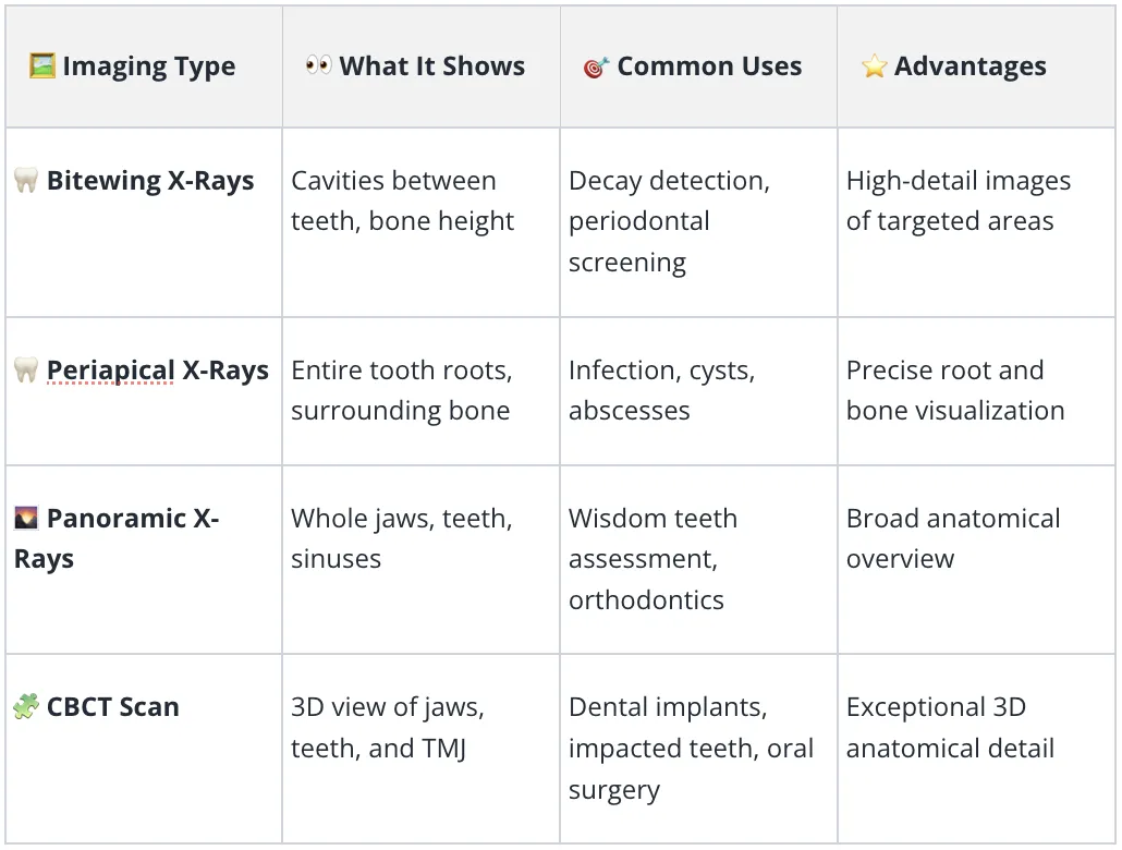
Bitewing Dental X-Rays
Ideal for detecting early cavities between teeth and checking bone levels for gum disease.
Bitewing X-rays show the upper and lower back teeth (usually molars and premolars) on one small film or sensor, capturing where cavities often hide—between the teeth—and revealing early signs of bone loss from gum disease. These are typically recommended during regular dental exams to help diagnose and treat decay before it becomes visible or painful.
Periapical Dental X-Rays
Reveal tooth roots, surrounding bone, abscesses, cysts, and infections below the gumline.
Periapical X-rays focus on one or a few teeth, showing the entire length from crown to root tip and the bone supporting the teeth. That is essential for diagnosing root infections, abscesses, cysts, or bone changes that may require root canal therapy or further dental care.
Panoramic Dental X-Rays
Capture a single image of the upper and lower jaws, helpful for viewing developing or impacted teeth and for orthodontic and oral surgery planning.
Panoramic X-rays offer a broad "big picture" view—imaging all the teeth, jawbones, and important structures in a single sweep. They’re commonly used for planning orthodontic treatments, evaluating wisdom teeth, screening for jaw cysts or tumors, and assessing bone structure for dental implants or extractions.
CBCT Scan (Cone Beam Computed Tomography)
Uses a cone-shaped X-ray beam to render detailed 3D images of oral structures, teeth, bone, and nerve pathways.
Essential in diagnostic dentistry for complex cases needing precise anatomical data.
CBCT scans are a form of advanced 3D dental imaging that uses a cone-shaped beam computed tomography machine to capture a detailed model of your teeth, jaws, nerves, and sinuses. This imaging method is especially valuable for complex procedures—such as dental implant planning, evaluating impacted teeth, or preparing for oral surgery—where a standard 2D dental X-ray may not provide enough information.
CBCT Scan: What Is Cone Beam Computed Tomography in Dental Imaging?
A CBCT scan uses cone beam computed tomography to create 3D views of the teeth, bone, nerves, and soft tissues, surpassing the two-dimensional images from traditional dental X-rays.
With CBCT, dental radiographs reveal complex anatomy crucial for treatment planning in diagnostic dentistry.
Unlike standard dental X-rays, which create flat, two-dimensional pictures, CBCT (cone beam computed tomography) provides a detailed three-dimensional (3D) model of your mouth and jaws. During a CBCT scan, a special machine rotates around your head, capturing hundreds of thin image "slices" from every angle. Advanced computer software then compiles these slices into a 3D image that helps dentists see not only teeth and jawbones, but also nerves, roots, sinuses, and airways.
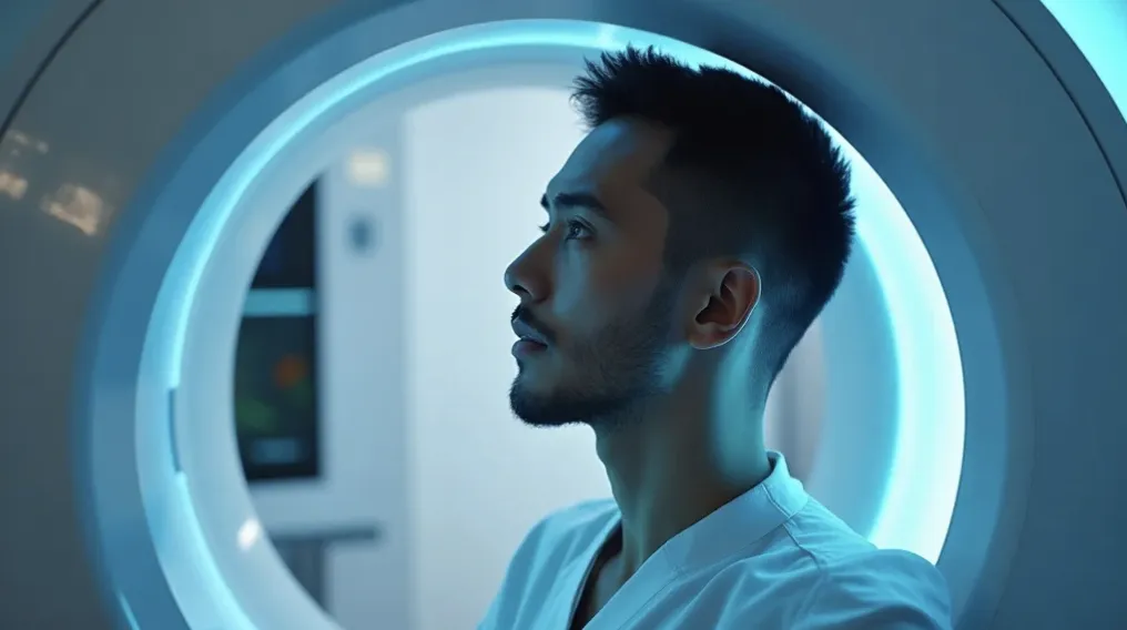
CBCT scans are prescribed when conventional dental X-rays can't provide key anatomical details needed for safe, effective treatment planning.
What is the difference between dental X-rays and CBCT?
Dental X-rays are two-dimensional images best for routine diagnosis, while CBCT provides 3D imaging for advanced dental exams, such as planning implants or evaluating bone and root structures.
The essential difference is detail and dimension. Traditional dental X-rays are fast, convenient, and provide enough information for most regular exams and basic treatment. CBCT scans, on the other hand, deliver high-resolution 3D data, making it possible to see complex root shapes, nerve locations, bone thickness, and abnormal growths from every angle—critical information for dental implants, oral surgery, and challenging root canal cases. CBCT is generally reserved for situations where 2D imaging isn’t enough.
When Is CBCT Recommended for Dental Patients?
Dental implant planning: Accurate assessment of bone structure and nerve locations.
Identifying impacted teeth that are not visible on standard dental X-rays.
Complex root canal evaluation for teeth with unusual anatomy or previous failures.
Oral surgery planning, including extractions, cysts, or tumors.
Orthodontic assessment when a panoramic or cephalometric X-ray does not provide enough detail.
TMJ evaluation to identify bone abnormalities or joint disorders.
Each CBCT recommendation is based on clinical justification; not every patient needs 3D dental imaging.
A CBCT scan is not part of every dental visit. Instead, this imaging procedure is targeted for cases where conventional dental radiographs cannot clearly show the full anatomy or problem area. If you’re considering a dental implant, CBCT maps bone volume, nerve pathways, and sinus positions—helping your dentist choose the best implant size, angle, and placement while minimizing risk. For impacted or extra teeth (especially wisdom teeth), CBCT imaging clarifies the neighboring roots and nerves, which improves safety during extraction.
For root canal therapy, CBCT provides intricate views of root canal shapes, fracture lines, or previous treatment. In orthodontics or with TMJ issues, it can reveal nuances in bone or joint structures that 2D X-rays might miss. Not all patients will ever need CBCT. Dentists recommend it only when the additional information changes diagnosis or treatment. This careful, evidence-backed approach is highlighted by the American Dental Association and the American Academy of Oral and Maxillofacial Radiology.
Dental X-Ray Safety and CBCT Scan Radiation Exposure
Modern dental X-rays involve very low levels of radiation exposure due to digital technology and strict safety guidelines.
Cone beam computed tomography emits more radiation than a single dental X-ray, but is much lower than conventional medical CT scans.
Dentists follow safety protocols and the ALARA (As Low As Reasonably Achievable) principle to minimize patient risk.
Dental imaging is performed only when there is a clear diagnostic or treatment benefit.
Every imaging exam—dental X-ray or CBCT scan—uses ionizing radiation, which carries a small risk. Dental X-rays are designed to use the lowest possible amount of radiation for a clear picture, and digital sensors require even less than traditional film. For context, the exposure to radiation from modern dental radiographs is very low; for most people, it equals only a tiny fraction of the naturally occurring “background” ionizing radiation experienced in daily life.
Every dental X-ray and CBCT scan is prescribed based on your individual oral health needs—not as a routine procedure.
CBCT scans generate higher radiation than a single dental X-ray, but still much less than hospital-grade medical CT scans. Dentists and dental staff rigorously follow safety guidelines and the “ALARA” principle—using radiation only As Low As Reasonably Achievable—balancing diagnostic benefit with minimal risk. Safety equipment, such as lead aprons and thyroid collars, is often used—especially for children and those at higher risk.
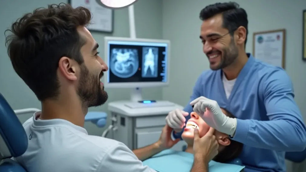
It’s important to remember that every dentist’s decision to recommend an X-ray or CBCT scan is tailored: weighing your age, dental history, symptoms, and needs. The goal is not only accurate diagnosis but also maximum safety and peace of mind. If you have concerns about radiation exposure or safety, never hesitate to ask your dental team—they welcome your questions and will help you make an informed choice.
What Happens During a CBCT Scan? Step-by-Step Patient Guide
Preparation: You may remove metal jewelry or glasses for clear images.
Positioning: You’ll stand or sit as the dental imaging machine positions your head securely.
Image capture: The CBCT scanner slowly rotates around your head for 20–40 seconds.
Image review: Your dentist reviews high-resolution 3D dental images for diagnosis and treatment planning.
The CBCT experience is straightforward and comfortable. There are no shots, no pain, and no need to bite on sensors. Instead, your dentist or technician will help you remove any metal items, like glasses or earrings, and explain the process. You stand or sit, resting your chin or forehead on the scanner support. The cone beam computed tomography machine then rotates smoothly around your head, taking detailed digital images from every angle as you remain still. You’ll hear a gentle hum but feel nothing. The scanning itself takes less than a minute. Once complete, your dental team will digitally “assemble” the images into a precise 3D model, ready for review and careful treatment planning.
Benefits and Limitations of Dental X-Rays and CBCT Imaging
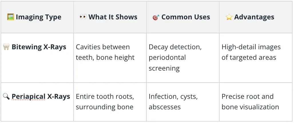
Dental imaging can detect decay, infection, cysts, fractures, bone loss, impacted teeth, and some tumors.
Radiographs cannot replace a thorough dental exam. Not all dental conditions require X-rays or CBCT scans.
Imaging complements—not replaces—professional dental judgment and clinical findings.
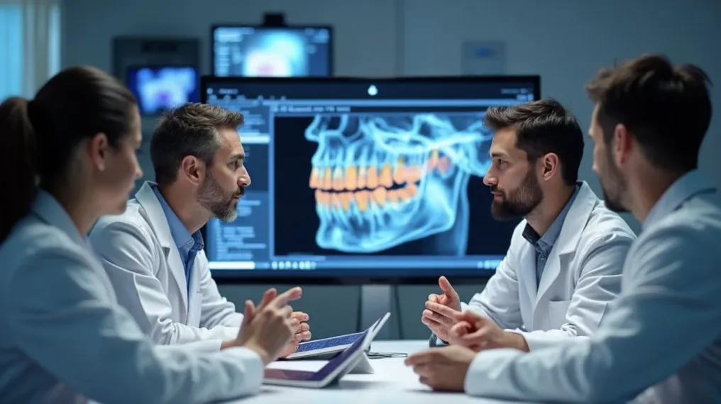
Dental X-rays and CBCT scans play a supporting role in oral health diagnosis—giving your dentist a more complete picture, but never replacing their expertise or a hands-on dental exam. While these imaging procedures detect cavities, bone loss, infections, fractures, impacted teeth, and even some tumors, not all dental conditions are visible on a radiograph. Some issues are best found through clinical signs, patient symptoms, or a combination of both. Trust your dental team’s experience and expect them to explain when (and why) any particular imaging study is recommended.
People Also Ask about Dental X-Rays and CBCT Scans
What are the three types of dental X-rays?
The main dental X-rays are bitewing, periapical, and panoramic. Each serves different diagnostic purposes in a dental exam.
Bitewing X-rays target the areas between back teeth, ideal for spotting cavities and assessing bone levels. Periapical X-rays capture the entire tooth root and surrounding bone, helpful for diagnosing root infections and abscesses. Panoramic X-rays show a wide view of both jaws, perfect for wisdom teeth, bone structure, orthodontic planning, and oral surgery cases.
What are the new guidelines for dental X-rays?
Dentists follow evidence-based guidelines, using dental radiographs only when needed based on age, symptoms, and risk factors, not on a fixed schedule.
National guidelines—such as from the American Dental Association—emphasize that dental X-rays should never be one-size-fits-all. Dentists base imaging decisions on your oral health, history, and risk factors. Radiographic exams are recommended only if the expected benefit outweighs any risk, and the choice is tailored for every patient, every time.
Should I decline dental X-rays?
Dental X-rays are recommended only when clinically justified. Discuss any concerns with your dentist, who can explain risks, benefits, and alternatives.
It’s natural to be cautious about radiation. However, dental X-rays are prescribed only when there’s a benefit for your diagnosis or care. If you’re worried, talk with your dental team—they can explain how your exam is tailored to minimize risks and only performed when truly necessary for your oral health.
How often does an adult need dental X-rays?
Frequency depends on oral health, age, dental history, and risk for cavities or gum disease. Some adults need X-rays every 1–2 years, others less often.
There’s no universal schedule: some adults may need X-rays each year if they have a high risk of cavities or gum disease, while others—especially those with healthy teeth and minimal dental work—might only need them every few years. Trust your dentist to recommend the right timing based on your unique needs.

Frequently Asked Questions: Expert Answers About Dental X-Rays and CBCT Scans
What are dental X-rays?
Dental X-rays are images created using low-dose radiation to show the teeth, jaws, and supporting structures. These radiographs help dentists spot cavities, infections, bone loss, and abnormalities that cannot be seen with a regular dental exam.
What is cone beam computed tomography (CBCT)?
CBCT is a specialized dental imaging technique that provides three-dimensional (3D) X-ray images of the mouth, jaws, and facial structures. It offers detailed anatomical information, essential for complex cases such as dental implant planning and oral surgery.
Why would my dentist recommend CBCT?
A dentist recommends CBCT when conventional dental X-rays don’t provide enough information, such as for planning dental implants, assessing impacted teeth, evaluating TMJ disorders, or investigating unexplained pain or swelling.
Is CBCT better than traditional X-rays?
CBCT isn’t always better—it's different. It is invaluable for complex diagnoses requiring 3D detail but isn’t necessary for common issues that 2D dental X-rays can clearly diagnose.
How safe are dental X-rays?
Dental X-rays use minimal radiation, especially with digital advances. Dentists recommend them only when the value of the information gained outweighs the potential risks. X-rays are not performed routinely without clinical need.
How long does a CBCT scan take?
A CBCT scan typically takes less than one minute. Preparation and explanation usually add 5–10 minutes to your appointment, after which your dentist will review the 3D images.
What conditions can CBCT diagnose?
CBCT can detect anatomical issues such as extra teeth, impacted teeth, jaw tumors, bone defects, TMJ disorders, and provide detailed data for dental implant planning and root canal assessment.
How should I prepare for CBCT?
Typically, preparation involves removing metal objects from your head and neck, such as jewelry. Always let your dentist know if you are pregnant or might be pregnant.
Can children have CBCT imaging?
CBCT imaging is generally reserved for children only when traditional dental X-rays do not provide enough information to ensure proper diagnosis and treatment.
How often are dental X-rays needed?
There is no one-size-fits-all schedule. Your dentist will consider your oral health, risk of disease, age, and history before recommending dental imaging at appropriate intervals.
Our team is dedicated to your oral health and safety. If you have any questions about dental X-rays, CBCT scans, or want a personalized imaging plan, contact us today to find a trusted dental care provider in your community.
Understanding dental X-rays and CBCT scans is just one part of making informed decisions about your oral health. If you’re ready to take the next step—whether it’s finding a dentist who prioritizes advanced diagnostics or exploring comprehensive care options—consider browsing the dental directory for a curated list of experienced professionals. This resource can help you connect with providers who offer the latest in dental technology and patient-centered care. Empower yourself with knowledge and access to the right dental team, so you can approach every appointment with confidence and clarity.
S. Raj, M.D. / Bestdentists.com
Director, Search Intelligence University/IABLEd
International Association for Business Leaders’ Education
Sources
https://www.ada.org/resources/ada-library/oral-health-topics/x-rays-radiographs - X-Rays/Radiographs
https://www.deltadental.com/protect-my-smile/procedures/x-rays/ - Dental X-rays: Key Things to Know

The IABLEd Dental Intelligence University
The Definitive Curriculum for Modern Dentistry and Oral Health
Praevenire. Curare. Sorridere.
Prevent. Treat. Smile.
Want Your World to Discover You,
the Easy Way?
Experience exceptional dental care while discovering the confidence of a healthier smile.
We provide the expertise, technology, and personalized treatment to help you achieve lasting oral health and beautiful, confident smiles. Ask us how.
Copyright 2026. Best Dentist Near Me. All Rights Reserved.


* * * * *
COMPANY
30721 Russell Ranch Rd Suite 140, Westlake Village, CA 91362
CUSTOMER CARE
Get In Touch
Mon – Thurs
10:00am – 4:00pm
Fridays by Appointment
Saturday, Golf
Sunday –hurch
LEGAL
Stay informed about how new technology can make or break a brand.
FOLLOW US


Prevent. Restore. Smile.
© 2026 IABLEd Dental Intelligence Academy | All Rights Reserved.
1: Oral Health Foundations 2: Preventive Dentistry & Patient Care 3: Restorative Dentistry & Treatment Planning 4: Advanced Clinical Techniques & Digital Dentistry 5: Cosmetic & Comprehensive Dental Care 6: Bonus Dental Masterclass Series